In genome modification processes, nucleases play a key role. Thanks to their ability to cleave DNA at or near specific recognition sites, they allow us to specifically cut (and then paste) our genetic material. Among all the different enzymes out there, an interesting example is that of I-SceI, as several studies have recently focused on this endonuclease and its possible applications for Pseudomonas putida genome engineering.
Pseudomonas putida is a very particular microorganism. It presents a versatile metabolism that allows it to degrade organic solvents such as toluene. In addition, these organisms can be genetically modified to function as organic-compound production systems for agricultural and pharmacological purposes. Despite the many practical applications Pseudomonas putida has, there is a lack of synthetic biology tools specific for non-conventional organisms like putida.
Recently, more and more tools for Pseudomonas’ genetic modification are developing. As some of these studies have used SEVA plasmids in their experiments, here we review two of them that show two promising features of I-Scel: plasmid curing and accelerated genome engineering.
Easy target- and self-curing of vectors to save time in genome modification
Pseudomonas genome editing is usually based on homologous recombination regulated by DNA-modifying enzymes encoded by plasmids. However, once the heterologous genes are expressed and the modification has taken place, it may be useful to eliminate those plasmids, using a process often referred to as plasmid curing. This step can be tedious and time-consuming, and most methods can be impractical when applied to P. putida.
In this article [1], researchers from the SEM Center at the Technical University of Denmark have enabled plasmid-curing in a simple way based on in vivo digestion of the vectors by the I-SceI homing nuclease, that can induce double strand breaks into plasmids thus putting an end to their replication. The process is controlled using a chemical inducer: 3-methylbenzoate.
SEVA plasmids can be modified by inserting an I-SceI recognition site into a conserved region present in all of them within the antibiotic resistance marker and the oriT module. A one-step, site-directed PCR mutagenesis of any SEVA vector can be used to this end! Using this method, 3-methylbenzoate can induce plasmid cleavage easily, therefore enabling plasmid curing when using SEVA plasmids for P. putida genome modification.
Accelerated engineering by recombination and CRISPR-aided counterselection
Recently, an increase in the use for bioproduction of Pseudomonas species has been observed, as a consequence of their specific characteristics. However, most synthetic biology tools developed so far are only applicable to obtain certain types of mutations or can’t be used in several Pseudomonas species.
The SEM Center team have developed a system [2] based on DNA recombination forced by the formation of double strand breaks using I-SceI meganucelase followed by counterselection of mutants aided by a synthetic CRISPR-Cas9 device. Several experiments were required to refine this process and most of them were developed using SEVA plasmids. Finally, this protocol was established, combining different molecular elements of the toolbox and creating a quick and smooth workflow for genome edition of P. putida.
These studies demonstrate the versatility of SEVA plasmids for different purposes. The expansion of this standard to more laboratories worldwide could be key to facilitating advances in different areas of research, such as synthetic biology.
References
[1] Volke, Daniel C., et al. «Synthetic control of plasmid replication enables target-and self-curing of vectors and expedites genome engineering of Pseudomonas putida.» Metabolic engineering communications 10 (2020): e00126.
[2] Wirth, Nicolas T., Ekaterina Kozaeva, and Pablo I. Nikel. «Accelerated genome engineering of Pseudomonas putida by I‐SceI―mediated recombination and CRISPR‐Cas9 counterselection.» Microbial biotechnology 13.1 (2020): 233-249.
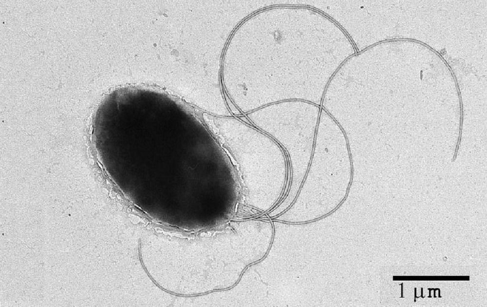
Featured photograph: Pseudomonas putida © CSIC